Mendgen kurt (8 Ergebnisse)

- Softcover
Anbieter: Clivia Mueller, Isernhagen, DeutschlandClivia Mueller
Verkäufer/-in kontaktierenVerkäufer/-in mit 5 SternenZustand: Gebraucht
EUR 6,26
EUR 24,10 VersandVersand von Deutschland nach USAAnzahl: 1 verfügbar
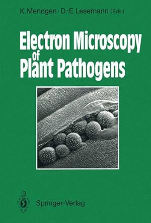
In den WarenkorbPlant Tissue(S.A.Arch.Microbiol.119)1978.S.113-117.Quart.m.zahlr.Abb.br.-2) -Sonderabdruck-.
Anbieter: Clivia Mueller, Isernhagen, DeutschlandClivia Mueller
Verkäufer/-in kontaktierenVerkäufer/-in mit 5 SternenZustand: Gebraucht
EUR 6,20
EUR 24,10 VersandVersand von Deutschland nach USAAnzahl: 1 verfügbar
In den Warenkorbduring the bean rust infection process (S. A. Physiol. Plant Pathology 6) 1975. S. 275 - 282. m. zahlr. Abb. geklammert -2) -Sonderabdruck-.

Sprache: Englisch
Verlag: Springer-Verlag Berlin and Heidelberg GmbH & Co. K, 1990
- Hardcover
Anbieter: Cotswold Internet Books, Cheltenham, Vereinigtes KönigreichCotswold Internet Books
Verkäufer/-in kontaktierenVerkäufer/-in mit 5 SternenZustand: Gebraucht
EUR 90,54
EUR 16,41 VersandVersand von Vereinigtes Königreich nach USAAnzahl: 1 verfügbar
Text in English. Ex-university library with stamps on pastedowns and top/bottom page edges; remnant of label on rear free endpaper. Pages faintly yellowed but otherwise clean and tidy in tight binding. Used - Good. Good ex-lib hardback Used - Good. Good ex-lib hardback.

- Softcover
Anbieter: Ria Christie Collections, Uxbridge, Vereinigtes KönigreichRia Christie Collections
Verkäufer/-in kontaktierenVerkäufer/-in mit 5 SternenZustand: Neu
EUR 116,96
EUR 14,04 VersandVersand von Vereinigtes Königreich nach USAAnzahl: Mehr als 20 verfügbar
Zustand: New. In.

- Softcover
Anbieter: preigu, Osnabrück, Deutschlandpreigu
Verkäufer/-in kontaktierenVerkäufer/-in mit 5 SternenZustand: Neu
EUR 95,25
EUR 70,00 VersandVersand von Deutschland nach USAAnzahl: 5 verfügbar
Taschenbuch. Zustand: Neu. Electron Microscopy of Plant Pathogens | Kurt Mendgen (u. a.) | Taschenbuch | xv | Englisch | 2011 | Springer | EAN 9783642758201 | Verantwortliche Person für die EU: Springer Verlag GmbH, Tiergartenstr. 17, 69121 Heidelberg, juergen[dot]hartmann[at]springer[dot]com | Anbieter: preigu.
- Softcover
Anbieter: Revaluation Books, Exeter, Vereinigtes KönigreichRevaluation Books
Verkäufer/-in kontaktierenVerkäufer/-in mit 5 SternenZustand: Neu
EUR 156,83
EUR 14,65 VersandVersand von Vereinigtes Königreich nach USAAnzahl: 2 verfügbar
Paperback. Zustand: Brand New. 336 pages. 9.40x6.80x0.90 inches. In Stock.
- Softcover
Anbieter: AHA-BUCH GmbH, Einbeck, DeutschlandAHA-BUCH GmbH
Verkäufer/-in kontaktierenVerkäufer/-in mit 5 SternenZustand: Neu
EUR 106,99
EUR 63,05 VersandVersand von Deutschland nach USAAnzahl: 1 verfügbar
Taschenbuch. Zustand: Neu. Druck auf Anfrage Neuware - Printed after ordering - Plants, fungi, and viruses were among the first biological objects studied with an electron microscope. One of the two first instruments built by Siemens was used by Helmut Ruska, a brother of Ernst Ruska, the pioneer in constructing electron microsc…opes. H. Ruska published numerous papers on different biological objects in 1939. In one of these, the pictures by G. A. Kausche, E. Pfankuch, and H. Ruska of tobacco mosaic virus opened a new age in microscopy. The main problem was then as it still is today, to obtain an appropriate preparation of the specimen for observation in the electron microscope. Beam damage and specimen thickness were the first obstacles to be met. L. Marton in Brussels not only built his own instrument, but also made considerable progress in specimen preparation by introducing the impregnation of samples with heavy metals to obtain useful contrast. His pictures of the bird nest orchid root impregnated with osmium were revolutionary when published in 1934. It is not the place here to recall the different techniques which were developed in the subsequent years to attain the modern knowledge on the fine structure of plant cells and of different plant pathogens. The tremendous progress obtained with tobacco mosaic virus is reflected in the chapter by M. Wurtz on the fine structure of viruses in this Volume. New cytochemical and immunological techniques considerably surpass the morphological information obtained from the pathogens, especially at the host-parasite interface.
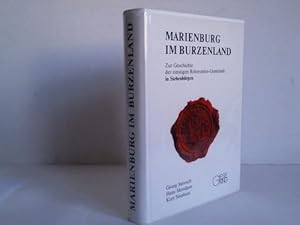
Marienburg im Burzenland. Zur Geschichte der einstigen Ritterorden-Gemeinde in Siebenbürgen
Siebenbürgen - Janesch/Troll, Georg / Mendgen, Hans / Stephani, Kurt
- Hardcover
Anbieter: Celler Versandantiquariat, Eicklingen, DeutschlandCeller Versandantiquariat
Verkäufer/-in kontaktierenVerkäufer/-in mit 5 SternenVerbandsmitglied: GIAQ
Zustand: Gebraucht
EUR 200,00
EUR 38,00 VersandVersand von Deutschland nach USAAnzahl: 1 verfügbar
2. Aufl. Gieseking Verl., Bielefeld, 1989. XII/456 S. mit einigen Tafeln u. 1 gefalt. Karte in der Anlage., Pbd. (Name auf Vorsatz)---- Gutes Exemplar - 1505 Gramm.


