Plasma Source Mass Spectrometry
Current Trends and Future Developments
By Grenville Holland, Dmitry BanduraThe Royal Society of Chemistry
Copyright © 2005 The Royal Society of Chemistry
All rights reserved.
ISBN: 978-0-85404-663-8Contents
Bio-analytical Applications,
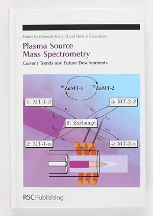
Metalloprotein Crosstalk: Quantifying Zinc Exchange Between Stable Isotopically Labeled Proteins Using Directly Coupled HPLC-ICP-MS A.Z. Mason, T.M. Potter, J. Webster and R.F. Meraz, 3,
Speciation of Selenium Compounds by Capillary Electrophoresis ICP-MS – Evaluation of ICP-DRC-MS Detection and Different Quantification Methods J. Nurmi, G. Koellensperger, G. Stingeder, D. Metze and N. Jakubowski, 28,
Application of ICPMS and Chromatography Based ICPMS to Studies of Metal-Protein Binding in Bioanalysis D. Gray, R. Burns, P. Brunswick, J. Le Huray, W. Chan, M. Mast, K. Allamneni and S. Sreedharan, 43,
Determination of Total Mercury in Urine By Inductively Coupled Plasma Mass Spectrometry (ICP-MS) P. Parsons, C. Palmer, K. Caldwell and R. Jones, 59,
Labile Arsenic Compounds in Biological Matrices, or Possible Problems Finding the Metal Species Present in Cells A. Raab, H. Hansen and J. Feldmann, 72,
Speciation Analysis with GC-ICP-MS: Organometal Detection in Tobacco Smoke M. Elobeid, Y. Chai, D. Clarke, R. Hannigan and J. Russ, 80,
Reaction Cells and High Resolution,
Determination of 44Ca-Isotope Markers for Parasitoid Studies Using Dynamic Reaction Cell ICPMS B. Hattendorf H. Wanner, H. Gu, S. Dorn and D. Gunther, 91,
Application of ICP-MS with a Hexapole Collision and Reaction Cell as an Element Selective Phosphorus Detector for Quality Control of Pharmaceuticals M. Edler, E. Busker, I. Feldmann, N. Jakubowski and C. Venzago, 99,
Advantages of Dynamic Reaction Cell ICP-MS for Geological Analyses K. Neubauer, S. Beres, L. Dionne and D. Green, 111,
Characterization and Production Ratio of Polyatomic Ion Interferences in Inductively Coupled Plasma Mass Spectrometry Using a High Resolution Mass Spectrometer Y. Takaku, D. Hinds and T. Shimamura, 120,
Measurement of Actinides at Ultra-Trace Level By Double-Focusing Sector-Field ICP-MS: Instrumental Performances and Analytical Difficulties F. Pointurier, N. Baglan and Ph. Hémet, 130,
Trends in Instrumentation,
Biomolecular and Atomic Mass Spectrometry: Good Friends or Uncaring Strangers? A. Makarov, 145,
Love Triangle: Trapping Fields, Ion Energy and Ion Molecule Reactions V. Baranov, D.R. Bandura and S. Tanner, 155,
Investigation of Vaporization of Laser Ablation Generated Aerosol and Monodisperse Particles in a Dry Ar ICP Using Time-Resolved Mass Spectrometry J. Olesik and N. Casey, 164,
Environmental Applications,
Signal Smoothing Device for Improving Analytical Precision in LA-ICP-MS and Its Applications A. Tunheng and T. Hirata, 177,
Metrology in Chemistry: ICP-Analysis in Environmental Science T. Prohaska, Y. Aregbe and P. Evans, 190,
ICP-MS as Analytical Tool on Spent Nuclear Fuel Analogues Studies J. Cáceres, J. Quiñones, E. Iglesias and A. Martinez-Esparza, 207,
Preliminary Work for the LA-ICP-MS Analysis of Carnelian Archaeological Artefacts S. Fraser, D. Polya, P. Lythgoe and T. Insoll, 213,
Selenium Speciation in Amphibian Larvae Developing in a Coal Fly Ash Settling Basin B. Jackson, W. Hopkins, J. Unrine, J. Baionno and T. Punshon, 225,
Speciation of Cancerostatic Platinum Compounds in a Waste Water Pilot Plant Zs. Stefánka, S. Hann, K. Lenz, G. Koellensperger, M. Fuerhacker and G. Stingeder, 235,
Multi-collectors,
Accurate Determination of Pt Concentrations at Ultra-low Levels (<<20Pg) in Clinical DNA Samples with High HF/PT Ratios Using the Thermo Finnigan Neptune Plasma Ionisation Multi-collector Mass Spectrometer (PIMMS) G. Nowell, D. Pearson, C. Ottley and M. Tilby, 245,
Novel Applications of MC-ICPMS for Isotope Dilution P. Evans, C. Wolff Briche, R. Hearn and T. Catterick, 259,
Solution and Laser Ablation Analysis of Sulphur Isotopes with the Neptune High Resolution Multi-collector ICP-MS (MC-ICP-MS): Application to Diffusive Gradients in Thin Films D. Bellis, G. Nowell, C. Ottley, D. Pearson and W. Davison, 268,
Accuracy and Precision in Plasma Ionisation Multi-collector Mass Spectrometry: Constraints from Neodymium and Hafnium Isotope Measurements D. Pearson and G. Nowell, 284,
Semiconductors,
Application of ICP-MS Technology to Semiconductor Manufacturing J.-M. Collard and Y. Kishi, 317,
Author Index, 334,
Subject Index, 337,
CHAPTER 1
Section 1
Bio-analytical Applications
METALLOPROTEIN CROSSTALK: QUANTIFYING ZINC EXCHANGE BETWEEN STABLE ISOTOPICALLY LABELED PROTEINS USING DIRECTLY COUPLED HPLC-ICP-MS
A.Z. Mason, T.M. Potter, J. Webster and R.F. Meraz
Department of Biological Sciences and Institute for Integrated Research in Materials, Environments and Society, California State University, Long Beach, 90840, USA
1 INTRODUCTION
While the importance of Zn as an essential but potentially toxic nutrient has been known for many years, it is only recently, since the completion of the human genome project, that we have come to appreciate just how many genes exhibit putative Zn binding motifs. Conservative estimates indicate perhaps a thousand or more human genes may be zinc regulated and, while we do not yet understand the functioning of many of these proteins, it is clear that Zn is an important cellular signal that can control many aspects of cellular functioning.
1.1 Metallothionein and Trace Metal Homeostasis
One protein that has been advocated to play a central role in intracellular metal metabolism and homeostasis in mammals and other organisms is metallothionein (MT). Metallothioneins (MT's) represent a super gene family of highly conserved proteins that have been implicated in the redox mediated distribution of Zn in animal cells. Four major isoforms of the protein have been identified in humans that are differentially expressed in various tissues. Two of the better-characterized isoforms, termed MT-1 and MT-2, typify the primary structure of the gene family in having a high cysteine content (30%) and no aromatic residues. It is the uniquely high composition of cysteine that accounts for the redox sensitivity of these proteins and their ability to co-ordinate seven Zn or Cd atoms. High resolution X-ray crystallographic studies have shown the protein to have a dumb-bell shape exhibiting 2 metal binding domains. The N-terminal β=domains of each isoform contain nine cysteines coordinating 3 zinc atoms while the C-terminal α-domains are comprised of 11 cysteines, coordinating 4 zinc atoms. The metals are bound tetrahedrally within these domains via thiolate bonds that either form bridges between adjacent metal atoms or act as terminal ligands for the metal. Although these metal-thiol clusters are thermodynamically stable (KD Zn = 1.2 x 10⁻¹³ M at pH 7.0), studies have shown that the metal atoms within the cluster are relatively labile, particularly in the β-domain, permitting both intramolecular and intermolecular transfer of Zn within and between given MT isoforms. This apparent lability, which can be increased by the presence of cellular oxidants, also accounts for the ability of the protein to donate Zn to the apo form of various metalloenzymes having a lower affinity for the metal.
1.2 Considerations for Quantifying Transfer of Zn Between Metal-binding Ligands
The recent realization of the importance of Zn in life processes has underscored the need for not only a better understanding of Zn homeostasis and the role of various MT isoforms in this process, but also the development of new techniques to quantitatively monitor the movement of the metal from different sources and sinks in the cell. To date, three different approaches have been used to study the ability of MT to donate Zn, namely radiometric analysis, enzymatic reporter assays and electrospray ionization (ESI) and matrix assisted laser desorption ionization (MALDI) mass spectrometry (MS). Collectively these procedures can provide quantitative and kinetic information on the role of MT in the transfer process providing that extreme care and appropriate controls are undertaken to account for contaminating sources of metal, including adventitious metals associated with other ligands in the reaction. However, perhaps the best approach for unequivocally identifying and quantifying the MT-derived Zn is to label the donor protein with an isotope that can be distinguished from other, extraneous sources of metal. This has been accomplished in the past by the use of 65Zn-radiolabeled MT. While gamma spectroscopy provides a highly precise and sensitive technique for metal quantification, accurate estimations of metal transfer require that the specific activity of the radiolabel on the various proteins be known before and after incubation. Radio and stable isotopic stoichiometry can be determined by using radiometric analysis in conjunction with a technique for total metal analysis, such as graphite furnace atomic absorption spectrophotometry (GFAAS) or inductively coupled plasma-optical emission spectroscopy/mass spectroscopy (ICP-OES/MS). Following incubation, exclusive MT to apo-enzyme Zn exchange can be confirmed by demonstrating identical specific activities for the recipient apo-protein after incubation as the MT prior to the reaction. These analyses require corrections to be made for counting efficiency and geometry, which can be time consuming and laborious. However, the biggest limitation in using 65Zn as a tracer in this context is that only one donor protein can be studied at a time. This negates the possibility of studying interactions involving multiple donor and recipient proteins and, for this reason, very little is known about the functional differences of the various MT isoforms in Zn homeostasis within the cell. Thus, although the two major isoforms, MT-1 and MT-2, are co-expressed in the same tissues at the same time by the same inducers, not much is known about their ability to exchange metals and act either as a common or independent Zn reservoir for metalloenzyme activation/inactivation.
1.3 Application of ICP-MS for Monitoring Zn Transfer Between Proteins
The following article describes a novel ICP-MS procedure that has the potential for quantifying Zn transfer between four different donor proteins while simultaneously allowing accurate assessments of contributions from contaminating sources to be taken into account. To unequivocally differentiate between the sources and sinks of metals in the transfer process, the procedure requires that the different donor proteins be differentially labeled with specific stable isotopes of Zn prior to incubation together. Following interaction, the proteins are separated and analyzed by directly coupled high performance liquid chromatography inductively coupled plasma mass spectrometry (HPLC-ICP-MS). Metals on the resolved protein species are quantified by post column flow injection analysis (FIA) of analyte standards of known isotopic abundance and concentration. By using multiple recipient and donor proteins having different stable isotopic signatures, this technique has the potential for empirically studying and modeling the kinetic and thermodynamic aspects of Zn transfer between a number of competing ligands in relatively complex samples comparable to the cytoplasmic milieu. This is an important step to understanding the regulatory role of this metal in protein functioning and cellular metabolism.
To demonstrate the potential of this analytical method for evaluating new hypothesis about the mechanisms of metal exchange between metal-requiring proteins we have undertaken a number of clearly defined experiments using well characterized metalloprotein mixtures under non-physiological conditions. In the current study the procedure has been used to better understand the function and specific transfer properties of the two major MT isoforms (MT-1 and MT-2) and their ability to act as independent vectors in Zn transfer. More specifically, this paper describes the use of this procedure to study and model the time dependent intermolecular transfer of Zn between the two major MT isoforms, the relative transfer of Zn from the two isoforms to metal-depleted Alkaline Phosphatase (apo-AP), and the effect of apo-AP on the intermolecular transfer between the isoforms. To the best of our knowledge this is the first attempt to directly monitor inter- protein exchange of a metal by coupled HPLC-ICP-MS using two monoisotopically labeled proteins.
2 MATERIALS AND METHODS
2.1 Reagents and Materials
Rabbit liver metallothionein 1 and 2, bovine intestinal mucosa alkaline phosphatase Type VII-S, p-nitrophenyl phosphate (p-NPP), reduced glutathione (GSH), dithiothreitol (DTT), pyridine-2,6-dicarboxylic acid (PDC), 5',5'-dithiobis(2-nitrobenzoic acid) (DTNB: Ellman's reagent), acetonitrile, trifluoroacetic acid (TFA), Trizma® hydrochloride and base (Ultra-Pure) were purchased either from Sigma Chemicals or from Aldrich. Stable isotopic 67Zn and 70Zn were purchased from Oak Ridge National Laboratories. For the 67Zn, the isotopic abundances of 64Zn, 66Zn, 67Zn, 68Zn, and 70Zn were certified to be 1.113, 1.95, 94.6, 2.28 and 0.054 atomic % respectively with a precision of +/- 0.03% or better for all isotopes. For 70Zn, double pass isotope was used with a certified isotopic 64Zn, 66Zn, 67Zn, 68Zn, and 70Zn abundance composition of 0.13, 0.07, 0.02, 0.06 and 99.72 atomic % respectively with a precision of +/- 0.03% or better for all isotopes. Multi-element standards containing 10µg.mL-1 Zn, Cu, Cd, Ag and a single-element standard solution for Hg were obtained from SCP Science (Baie D'Urfé, Québec Canada, H9X 4B6). Each element was certified at >99.99% purity. Solutions were prepared in acid washed, plastic ware using Millipore Milli-Q® deionized water with a resistivity of >18MΩxcm-1. Adventitious metals in buffers and other preparative solutions were removed by filtration through a 2 x 40cm column packed with rejuvenated Chelex 100 beads (BioRad) at a rate of 1mlxmin-1. All solutions were checked for metal content by ICP-MS prior to use and rechelated if the Zn, Cu or Cd concentrations in the solution exceeded 0.05ng.mL-1.
2.2 Coupled HPLC-ICP-MS Instrumentation and Instrumental Parameters
HPLC was performed at a flow rate of 1ml.min-1 using a "biocompatible" Beckman 126 programmable solvent delivery module equipped with a Beckman 168 diode array UV/VIS detector. PEEK® tubing was used to connect the various components in the system. A scavenger column (7.8mm ID x 75mm length), hand-packed with Chelex 100 resin (BioRad), was installed prior to a 100µL PEEK® sample injector loop to remove metals from the solvent stream. The system was flushed for 10 minutes with 10% HNO3 to rejuvenate the scavenger column and passivate the system hardware. The system was subsequently flushed with deionized water and then the mobile phase before the attachment of the various size exclusion (SE) and weak anion exchange (AE) analytical columns.
Samples for directly coupled HPLC-ICP-MS were fractionated either isocratically by SE HPLC (600 x 7.8mm Biosep-SEC-S2000 column, Phenomenex) using a mobile phase of filtered, degassed 20mM Tris (pH 7.2) or by AE HPLC (75 x 7.8mm Showdex DEAE-825, column Phenomenex). A stepped gradient of 10mM Tris (pH 8.2) increasing to 10mM Tris in 500mM NH4Cl (pH 8.2) over a 60-minute period was used to fractionate the proteins from the AE column. The UV/VIS absorbance of the eluting proteins was monitored between 190 and 600nm. Metals associated with the proteins in the solvent stream were analyzed using a PE-6100DRC ICP-MS directly coupled to the HPLC. A Rheodyne 7125 injector with a 200µL flow injection loop installed immediately prior to the ICP-MS was used to quantify the elemental composition of the resolved peaks and monitor analyte recovery. Sample aspiration into the ICP-MS was through a quartz Meinhard concentric nebulizer and a cyclonic spray chamber. The instrument was tuned daily using a 10ng.mL-1 solution of Li, Mg, Ce, Co, In, Ba and U to minimize oxide adduct formation and doubly charged species without unduly compromising sensitivity. Count rates were typically better than 600 x 103 cps for 115In with a RSD of <0.5%. The Ce:CeO ratio was typically <0.02 under these operating conditions. The instrument was run in Non-DRC (Dynamic Reaction Cell) mode. Analysis and integration of the isotopic masses on the various peaks was performed using the Perkin Elmer Chromelink™ chromatographic application within Totalchrome™ software.
Preparation of Apo-Metallothionein (Thionein)
Thionein 1 and 2 were prepared by reverse phase HPLC using a modification of the method previously described by Vasak. In brief, approximately 0.1 mg of lyophilized Cd/Zn MT-1 or MT-2 was dissolved in 100µL of a 0.1% TFA in 15% acetonitrile (pH 2.0). The sample was then further acidified by the addition of 10µL of 100% (TFA). Following 10 minutes of incubation, the released free metal was separated from the thionein by reverse phase chromatography. For chromatography, 100 µL of the crude acidified sample (0.1667mM MT) was applied to a Jupiter™ C4 RP-300 column (5µ, 4.6 x 250mm; Phenomenex) and eluted at a flow rate 0.5ml/min using a stepped gradient increasing from 75% Buffer A (0.1% TFA, pH 2.0) to 100% Buffer B (0.1% TFA, pH 2.0 in 60% acetonitrile) over 60 minutes. The eluent was monitored continuously using a Beckman 168 photodiode array detector set to simultaneous record absorbances at 220nm (peptide bond), 254nm (Cd-thiolate bond) and 280nm (aromatic residues). The success of the procedure was monitored by the disappearance of the 254nm signature for the metal thiolate cluster coincident with the appearance of a peak showing strong absorbance at 220nm but not 280nm. Fractions conforming to these spectral criteria were collected by hand and the presence of free thiols was quantified spectroscopically at 412nm by the reductive cleavage of DTNB. A standard curve of GSH was used to quantify the thiol content of the thionein assuming a thiol stoichiometry of 21:1 between thionein and GSH. The thiol content was normalized to the thionein protein concentration which was determined spectroscopically at 220nm in 0.1M HCl using a extinction coefficient of 48,200 M-1cm-1. Thiol:apo-MT ratios of between 18-22 were typically obtained from the fractions as reported by others.
(Continues...)Excerpted from Plasma Source Mass Spectrometry by Grenville Holland, Dmitry Bandura. Copyright © 2005 The Royal Society of Chemistry. Excerpted by permission of The Royal Society of Chemistry.
All rights reserved. No part of this excerpt may be reproduced or reprinted without permission in writing from the publisher.
Excerpts are provided by Dial-A-Book Inc. solely for the personal use of visitors to this web site.