
Verwandte Artikel zu Teaching Atlas of Chest Imaging
Inhaltsangabe
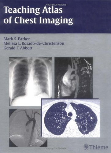
This lavishly illustrated book is your comprehensive, hands-on guide to evaluating chest images. It is ideal for reading cover-to-cover, or as a reference of radiological presentations for common thoracic disorders. With this book, you will learn to interpret chest images and recognize the imaging findings, generate an appropriate differential diagnosis, and understand the underlying disease process. The atlas begins with a review of normal thoracic radiography, CT, and MR anatomy, and goes on to present cases on a wide range of congenital, traumatic, and acquired thoracic conditions. Each case is supported by a discussion of etiology, pathology, imaging findings, treatment, and prognosis in a concise, bullet format to give you a complete clinical overview of each disorder. More than 1,050 high-quality images demonstrate normal and pathologic findings, and complementary scans demonstrate additional imaging manifestations of disease entities. Residents, fellows, and general radiologists called upon to interpret chest images will find this easy-to-use book invaluable as a learning tool and reference. It is also a must for thoracic radiologists, pulmonary physicians, and thoracic surgeons who must read chest images - especially of challenging cases.
Die Inhaltsangabe kann sich auf eine andere Ausgabe dieses Titels beziehen.
Über die Autorin bzw. den Autor
Mark S. Parker, Melissa L. Rosado-de-Christenson, Gerald F. Abbott
Von der hinteren Coverseite
More than 1,000 state-of-the-art images allow for easy differentiation of normal and pathologic findings! From lung cancer screening, diagnosis, and staging, to critical care imaging and occupational lung disease, chest imaging remains one of the most complex subspecialties in diagnostic radiology. This book is a complete, hands-on guide to evaluating chest images. It is ideal for reading cover-to-cover, or as an illustrated reference of radiological manifestations of common thoracic disorders. With the Teaching Atlas of Chest Imaging, you will expand your chest imaging interpretation skills and learn to recognize abnormal findings, generate an appropriate differential diagnosis, and better understand the underlying disease process. The atlas begins with a review of normal chest radiographic, CT, and MR anatomy, and goes on to present cases that show a wide range of congenital, traumatic, and acquired thoracic conditions. Each case is supported by a discussion of the etiology of the disease, its underlying pathology, typical and unusual findings, treatment, and prognosis in a concise, bulleted format to provide a comprehensive overview of each disorder. Highlights: - More than 1,000 high-quality images demonstrate normal and pathologic findings, and their variations - Case-based approach exposes the reader to a broad spectrum of frequently encountered diseases and developmental lesions - Coverage of a variety of conditions including atelectasis, pulmonary infections, congenital lesions, neoplastic disorders, diffuse lung disease, and many more - Complementary images included in selected cases demonstrate additional radiological manifestations of disease entities or differential diagnostic considerations - Pearls and Pitfalls help you retain key anatomic and clinical concepts Residents, fellows, and general radiologists will find this easy-to-use book invaluable as a learning tool and reference. It is also a key resource for thoracic radiologists, pulmonary medicine physicians, and thoracic surgeons. Mark S. Parker, M.D., Associate Professor, Diagnostic Radiology, Thoracic Imaging Section, Medical College of Virginia Hospitals-VCU Health System, Richmond, Virginia. Melissa L. Rosado-de-Christenson, M.D., F.A.C.R., is Clinical Professor, Department of Radiology, The Ohio State University College of Medicine and Public Health, Columbus, Ohio, and Adjunct Professor of Radiology at the Uniformed Services University of the Health Sciences in Bethesda, Maryland. Gerald F. Abbott, M.D., is Associate Professor, Department of Diagnostic Imaging, Brown Medical School, and Director of Chest Radiology, Rhode Island Hospital, Providence, Rhode Island.
„Über diesen Titel“ kann sich auf eine andere Ausgabe dieses Titels beziehen.
Suchergebnisse für Teaching Atlas of Chest Imaging

Beispielbild für diese ISBN
Teaching Atlas of Chest Imaging
Verlag:
Thieme Medical Publishers, Incorporated, 2006
ISBN 10: 3131390212
ISBN 13: 9783131390219
Gebraucht
Hardcover
Anbieter: Better World Books, Mishawaka, IN, USA
Verkäuferbewertung 5 von 5 Sternen

Zustand: Good. Pages intact with minimal writing/highlighting. The binding may be loose and creased. Dust jackets/supplements are not included. Stock photo provided. Product includes identifying sticker. Better World Books: Buy Books. Do Good. Artikel-Nr. 11000587-20
Gebraucht kaufen
EUR 80,08
Versand gratis
Versand innerhalb von USA
Versand innerhalb von USA
Anzahl: 1 verfügbar