Verwandte Artikel zu Correlative Light and Electron MIcroscopy (Volume 111)...
Correlative Light and Electron MIcroscopy (Volume 111) (Methods in Cell Biology, Volume 111) - Hardcover

Inhaltsangabe
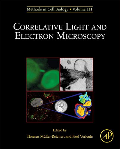
The combination of electron microscopy with transmitted light microscopy (termed correlative light and electron microscopy; CLEM) has been employed for decades to generate molecular identification that can be visualized by a dark, electron-dense precipitate. This new volume of Methods in Cell Biology covers many areas of CLEM, including a brief history and overview on CLEM methods, imaging of intermediate stages of meiotic spindle assembly in C. elegans embryos using CLEM, and capturing endocytic segregation events with HPF-CLEM.
Die Inhaltsangabe kann sich auf eine andere Ausgabe dieses Titels beziehen.
Über die Autorinnen und Autoren
Thomas Müller-Reichert is a Professor of Structural Cell Biology at the Technische Universität Dresden (TU Dresden, Germany). He is interested in how the microtubule cytoskeleton is modulated within cells to fulfill functions in mitosis, meiosis and abscission. The Müller-Reichert lab is mainly applying correlative light microscopy and electron tomography to study the 3D organization of microtubules in early embryos and meiocytes of the nematode Caenorhabditis elegans, and also in mammalian cells in culture. He has published over 75 papers and edited several volumes of the Methods in Cell Biology series on electron microscopy and CLEM.
TMR obtained his PhD at the Swiss Federal Institute of Technology (ETH) in Zurich and moved afterwards for a post-doc to the EMBL in Heidelberg (Germany). He was a visiting scientist with Dr. Kent McDonald (UC Berkeley, USA). Together with Paul Verkade, he set up the electron microscope facility at the newly founded Max Planck Institute of Molecular Cell Biology and Genetics (MPI-CBG). Since 2010 he is a scientific group leader and head of the Core Facility Cellular Imaging (CFCI) of the Faculty of Medicine Carl Gustav Carus of the TU Dresden. He acted as president of the German Society for Electron Microscopy (Deutsche Gesellschaft für Elektronenmikroskopie, DGE) from 2018 to 2019.
He taught numerous courses and workshops on high-pressure freezing and Correlative Light and Electron Microscopy.
Paul Verkade is a Professor of Bioimaging at the University of Bristol, UK where his research group works on the development and application of microscopy techniques to Biomedical questions. The main tools in the lab are Electron microscopy (EM) and Correlative Light Electron Microscopy (CLEM) in which fields he has published over 100 papers and edited 5 books on CLEM (including 4 Volumes of the Methods in Cell Biology series). PV obtained his PhD at the University of Utrecht, The Netherlands in 1996. Subsequently he did a post-doc at the EMBL, Heidelberg, Germany, after which he set up the electron microscopy unit at the newly formed Max Planck Institute for Molecular Cell Biology in Dresden, Germany from 2001. He moved to the UK in 2006 to set up another EM unit as part of an integrated LM and EM bioimaging facility, which facilitates CLEM workflows. He is actively involved in shaping the future microscopy landscape with roles in the Royal Microscopical Society and BioimagingUK and a current focus on putting volumeEM on the imaging map through community building and the organisation and co-chairing of the 1st Gordon Research Conference on vEM.
Von der hinteren Coverseite
The combination of electron microscopy with transmitted light microscopy (termed correlative light and electron microscopy; CLEM) has been employed for decades to generate molecular identification that can be visualized by a dark, electron-dense precipitate. This new volume of Methods in Cell Biology covers many areas of CLEM including a brief history and overview on CLEM methods, imaging of intermediate stages of meiotic spindle assembly in C. elegans embryos using CLEM, and capturing endocytic segregation events with HPF-CLEM.
„Über diesen Titel“ kann sich auf eine andere Ausgabe dieses Titels beziehen.
Neu kaufen
Diesen Artikel anzeigenEUR 7,44 für den Versand von Vereinigtes Königreich nach USA
Versandziele, Kosten & DauerSuchergebnisse für Correlative Light and Electron MIcroscopy (Volume 111)...
Correlative Light and Electron Microscopy
Anbieter: Majestic Books, Hounslow, Vereinigtes Königreich
Zustand: New. pp. 460 Illus. Artikel-Nr. 38598456
Anzahl: 3 verfügbar
Correlative Light and Electron Microscopy: Vol 111
Anbieter: Revaluation Books, Exeter, Vereinigtes Königreich
Hardcover. Zustand: Brand New. 1st edition. 404 pages. 9.50x7.75x1.25 inches. In Stock. Artikel-Nr. __0124160263
Anzahl: 2 verfügbar
